Architecture of Lost Space: Bio-Engineering and Transformations of the Maxillary Arch

An Analytical Reading in Osseous Morphology, Geographic Erosion, and the Master Planning of Facial Structural Systems
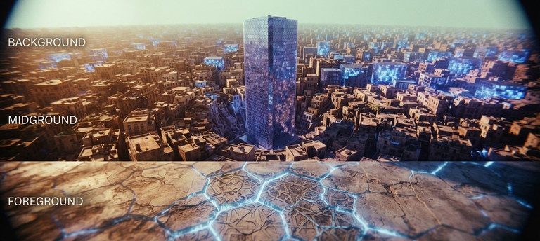
Biological infrastructure within the human body obeys laws similar to those governing urban fabrics and architectural configurations. When the primary columns supporting the structural grid—represented here by the teeth—are removed, the site does not cease to interact; rather, it begins reshaping its topography along inevitable vectors of erosion. The edentulous maxilla represents not merely an organic absence, but a profound spatial transformation that redraws the geographical map of the lower third of the face. Understanding this topographic metamorphosis and the volumetric retreat of alveolar bone redefines rehabilitative practice as a process of “spatial reconstruction” and “structural restoration” requiring meticulous site surveying, structural benchmark analysis, and a deep comprehension of the mechanical forces and directional regressions governing biological terrain.
The Primary Geometric Footprint: From Initial Plan to Spatial Contraction
The initial features of any biological structure are determined by its primary horizontal floor plan. In the case of the fully dentate maxillary arch, this initial geometry serves as a primary line of defense and load distribution. Foundational studies conducted by Nakatsuka and colleagues examining 62 dentate maxillary casts revealed that the natural arch form follows specific geometric diversity. The round-square pattern dominates in 58.1% of cases—subdivided into two branches based on the convergence or divergence of posterior components—followed by the square pattern at 21.0%, the purely round pattern at 4.8%, and finally the round V-shaped pattern at 16.1%. This native geometry constitutes the fundamental spatial code upon which the oral cavity is constructed.
However, once the dental supports are removed, this precise blueprint begins to yield and progressively alter. In a comprehensive microscopic study involving a morphological survey of 24 dry fully edentulous maxillary specimens, Pietrokovski and colleagues demonstrated that this complex geometric diversity contracts and shifts toward simpler forms as a result of erosion. Their findings showed that edentulous maxillary arches predominantly adopt an ovoid appearance in 65% of cases, while triangular forms retreat to 25%, and irregular configurations account for 10%. This transition from complex square and round layouts to ovoid and triangular footprints reflects the gradual collapse of the jaw’s peripheral zones, confronting the bio-architect or prosthodontist with a site operating under continuous areal contraction dynamics.
Dynamics of Centripetal Contraction: Vectors of Erosion and Topographic Collapse
Bone resorption following tooth extraction is not merely a reduction in elevation, but a spatially directed movement of warping and contraction. In a landmark classification that established a foundation for understanding jaw geography, Cawood and Howell analyzed 300 dry skulls, establishing a system to classify the stages of jaw degradation across six classes, ranging from the fully dentate jaw (Class I) to severe atrophy and basal bone erosion (Class VI). They demonstrated that while the basal bone structure maintains relative stability, the alveolar process—acting as the curtain wall surrounding the teeth—undergoes the vast majority of vertical and horizontal dimensional changes.
The most compelling aspect of this erosive mechanics lies in the directional discrepancy between the maxilla and mandible. Pietrokovski and colleagues established that bone resorption in the maxilla follows a centripetal and apical vector, meaning the residual ridge recedes inward toward the palate and upward. This morphological behavior stems from the fact that the structural basal bone of the maxilla lies internal to the original tooth placement. Consequently, the edentulous maxillary arch becomes narrower and shorter relative to its original footprint.
Conversely, mandibular resorption follows a centrifugal and apical trajectory, causing the mandibular arch to widen in appearance. This clash in contraction vectors creates a spatial architectural dilemma characterized by a reverse projection or direct edge-to-edge relationship, where the upper crest sits at the same level as, or internal to, the lower crest, confounding tectonic load-bearing axes.
Quantifying this spatial breakdown, Mercier and Lafontant introduced a quantitative system to evaluate residual ridge atrophy by measuring sagittal area in square millimeters on lateral cephalograms, dividing atrophy into five distinct tiers from absent to extremely severe. Their research revealed that the rate of area loss in the mandible exceeds that of the maxilla by a factor of four to one, while also demonstrating that female skeletal structures experience more severe atrophy than male counterparts, necessitating the inclusion of biological and gender factors when designing alternative rehabilitative structures.
Surveying Micro-Topography: Tomographic Scanning and Load-Bearing Site Capacity
Just as a structural engineer requires soil testing and three-dimensional topographic surveying prior to laying foundations, working with the maxillary arch demands a precise assessment of remaining bone thickness at the implant platform level. In this context, Bulyalert and Pimkhaokham introduced a modern classification of anterior alveolar arches based on cone-beam computed tomography (CBCT) by evaluating 113 tomographic images. Using the root centers of anterior teeth 3 millimeters below the cementoenamel junction as geographic reference points, they categorized arch forms into four fundamental types based on intercanine width, interpremolar width, and intercanine width-to-depth ratio.
These types include the long narrow form, characterized by severely limited transverse and anteroposterior dimensions that impose strict spatial constraints; the short medium form, exhibiting balanced and moderate dimensions in both directions; the long medium form, possessing moderate width alongside increased spatial depth; and the long wide form, representing the maximum footprint in both width and area depth.
The structural significance of this tomographic classification lies in revealing explicit variance in buccolingual bone thickness across these arch forms. Arches exhibiting the long wide configuration demonstrated the highest structural efficiency and bone thickness, averaging 9.26 millimeters at 3 millimeters below the cementoenamel junction and 9.88 millimeters at mid-root. Conversely, long narrow and short medium arches displayed the thinnest bone profiles. This morphometric data provides critical information for the bio-designer, as slender arches demand extreme caution when selecting structural fixture diameters and require preliminary bone augmentation procedures to expand the site prior to applying mechanical loads.
Benchmarks and Mathematical Modeling: Topographic Inference via Parabolic Curves
When surface features of a site degrade, surveyors rely on fixed benchmarks to reconstruct original maps. In the edentulous maxilla, the incisive papilla, labial and buccal frenula, maxillary tuberosity, pterygomaxillary notch, palatine rugae, and palatine raphe emerge as critical anatomical landmarks that Lynn highlighted for their controlling influence in setting the boundaries of complete rehabilitative structures.
In an explicit effort to digitize oral geometry, Preti and colleagues studied 1,000 dental plaster casts of dentate maxillae, deriving a mathematical parabolic model to predict the geometric shape and size of the anterior arch based on the distance from the incisive papilla to the labial surface of each anterior tooth. This mathematical equation is defined by the formula:
y=9.720K−0.012x−0.024x2
where K represents a variable dimensional parameter. Crucially, the researchers demonstrated that interbuccal frenulum distance in edentulous patients serves as a practical field indicator for selecting the appropriate parabola size for tooth positioning, where a large distance (54.1 mm) denotes a wide arch, a medium distance (50.2 mm) indicates a medium arch, and a small distance (47.5 mm) signifies a narrow arch. This model provides an applied tool to reconstruct lost geometry with extreme precision.
However, reference benchmarks themselves are not entirely immune to the effects of time and erosion. Klemetti and colleagues revealed that the location of the incisive papilla shifts anteriorly in appearance with ongoing bone resorption and increased duration of edentulism, resulting from the flattening and contraction of the anterior ridge. Their study also demonstrated that remaining facial bone width in the canine region correlates strongly with both the duration of edentulism and the patient’s overall skeletal mineral status, with lower systemic bone mineral density accelerating ridge erosion and thinning.
Classification of Osseous Ridges: Knife-Edge Slopes and Infrastructure Degradation
The residual osseous ridge of the maxillary arch exhibits diverse topographic profiles, ranging from stable surfaces to steep inclines. Surveys by Pietrokovski and colleagues showed that 22% of residual maxillary alveolar ridges transform into knife-edged crests measuring less than 2 millimeters in width, while 78% assume a flat-rounded contour. These knife-edge ridges are unevenly distributed across the arch, peaking in the anterior incisor region at 47%, while decreasing to 10% in posterior premolar and molar regions.
At the macro-dimensional level, the edentulous maxilla exhibits width dimensions ranging between 39 and 60 millimeters (averaging 48 millimeters) and length dimensions between 40 and 54 millimeters (averaging 47 millimeters). Anatomical studies further demonstrate that the residual ridge crest lies external to the cranial base, except at the nasal spine and zygomatic process where the crest recedes internal to these structures.
To evaluate this morphological diversity from a clinical and applied perspective, Sharma and colleagues applied the classification system established by the American College of Prosthodontists to 87 edentulous patients. They found that Type A, representing a favorable and stable ridge with adequate vestibular depth, well-defined tuberosity, and hamular notch free of tori, was the most prevalent at 46%. Type B, characterized by reduced posterior buccal vestibular depth and diminished posterior landmark clarity, accounted for 41.4%. Type C, defined by loss of anterior labial vestibular depth, minimal resistance to movement, and hyperplastic tissue, appeared in 12.6% of cases, while Type D, involving severe total vestibular loss and prominent nasal spine, was not observed.
Statistical analysis in the study confirmed that residual ridge morphology correlates significantly with the duration of edentulism; patients living longer without teeth progressed toward compromised states such as Type C, whereas age and gender exhibited no direct effect on this morphological shift. Furthermore, users of removable prostheses frequently maintained Type A ridges, suggesting that moderate functional stimulation may slow osseous erosion and support soft tissue adaptation.
Structural Reconstruction: The Lip-Tooth-Ridge Framework and Resolving Interfacial Spatial Constraints
Rehabilitative planning for the maxilla extends beyond managing solid bone mass to regulating the external facade and its interface with surrounding tissues. In this context, Pollini and colleagues introduced the Lip-Tooth-Ridge classification system as a conceptual framework connecting lip mobility, targeted tooth position, and the geometric architecture of the alveolar ridge, establishing four clinical categories to guide structural intervention.
Under this framework, Class I represents minimal tissue deficiency, forming an ideal environment for fixed implant-supported crown-and-bridge restorations without requiring prosthetic tissue replacement. Class II involves a vertical bone defect requiring height management, while Class III presents a horizontal deficit compromising bone thickness and necessitating width augmentation. Class IV encompasses a severe combined defect uniting vertical and horizontal loss, dictating the use of removable prostheses equipped with a labial flange to restore missing tissue volume and facial support.
This classification integrates aesthetic risk factors, determined by smile-line exposure and lip dynamics, alongside structural risk parameters based on available prosthetic space. In doing so, it offers a navigational compass for determining prosthesis design—whether fixed or removable—as well as the required number and angulation of structural anchor points.
The edentulous maxillary arch provides a compelling model for bio-engineering applications, where intrinsic morphological traits interact with centripetal resorption vectors to produce a dynamic, evolving terrain. Achieving successful rehabilitation in this vital space requires the clinician to act as an architect—interpreting site transformations, relying on structural benchmarks, and selecting rehabilitative solutions that restore tectonic and aesthetic balance to the human face.
✦ ArchUp Editorial Insight
The structural alteration of the maxillary arch is the predictable spatial consequence of load-grid decommissioning. When primary dental supports are extracted, unmitigated biological resorption vectors—specifically centripetal and apical regression—reorganize the underlying foundation according to mechanical stress gradients, systemic bone density, and temporal duration. Standardized diagnostic classifications, from tomographic surveying to the Lip-Tooth-Ridge framework, function as risk-governance systems that dictate whether rehabilitative interventions require physical site expansion via bone grafts or spatial adaptation through prosthetic cantilevering. The resulting morphological shift from complex, load-distributing layouts to contracted ovoid footprints and knife-edge crests demonstrates that biological form strictly mirrors the withdrawal of structural forces. What presents externally as facial volume loss is the logical end-state of an unanchored site adapting to internal vector pressures.
References
- Pietrokovski, J., R. Starinsky, B. Arensburg, and I. Kaffe. “Morphologic Characteristics of Bony Edentulous Jaws.” Journal of Prosthodontics, 2007.
- Preti, G., P. Pera, and F. Bassi. “Prediction of the Shape and Size of the Maxillary Anterior Arch in Edentulous Patients.” Journal of Oral Rehabilitation, 1986.
- Bulyalert, A., and A. Pimkhaokham. “A Novel Classification of Anterior Alveolar Arch Forms and Alveolar Bone Thickness: A Cone-Beam Computed Tomography Study.” Imaging Science in Dentistry, 2018.
- Cawood, J. I., and R. A. Howell. “A Classification of the Edentulous Jaws.” International Journal of Oral and Maxillofacial Surgery, 1988.
- Mercier, P., and R. Lafontant. “Residual Alveolar Ridge Atrophy: Classification and Influence of Facial Morphology.” The Journal of Prosthetic Dentistry, 1979.
- Pollini, A., J. Goldberg, R. Mitrani, and D. Morton. “The Lip-Tooth-Ridge Classification: A Guidepost for Edentulous Maxillary Arches. Diagnosis, Risk Assessment, and Implant Treatment Indications.” The International Journal of Periodontics & Restorative Dentistry, 2017.
- Klemetti, E., L. Lassila, and V. Lassila. “Biometric Design of Complete Dentures Related to Residual Ridge Resorption.” The Journal of Prosthetic Dentistry, 1996.
- Sharma, R., A. Bhochhibhoya, B. Acharya, and S. B. Rana. “Clinical Evaluation of Residual Ridge Morphology of Maxillary Arch in Relation to Ageing and Length of Edentulism.” Journal of College of Medical Sciences-Nepal, 2019.
- Lynn, B. D. “The Significance of Anatomic Landmarks in Complete Denture Service.” The Journal of Prosthetic Dentistry, 1964.
- Nakatsuka, M., Y. Iwai, S. S. Jue, et al. “A Morphological Study on the Classification of Maxillary Dental Arches.” Okajimas Folia Anatomica Japonica, 2004.